The State of AI in Dentistry 2026: A Field Guide to the Tools

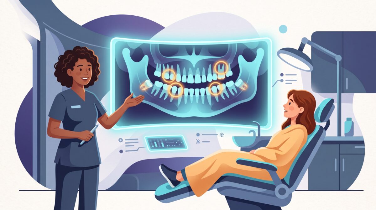
Here is something most people do not realize until they are already reclined in the chair: the AI hype cycle that swept through writing, coding, and customer service has quietly reached your dentist’s office too. Only in dentistry it did not arrive as a chatbot. It arrived as software that looks at your X-rays and points at things. And unlike a lot of AI, this kind is regulated, cleared, and increasingly ordinary. If your dentist has upgraded their imaging in the last couple of years, there is a real chance an AI has already read one of your radiographs.
This is the first piece in a series, so think of it as the map before the deep dives. No jargon dumps, no vendor cheerleading — just a plain-language field guide to what actually exists in 2026, what each kind of tool does, and what any of it means for you.
What “AI at the dentist” actually means now
Forget robots with drills. The AI showing up in dental offices today is almost entirely about reading images and organizing information. A dental X-ray is a surprisingly hard thing to interpret — findings are small, low-contrast, and easy to overlook at the end of a long day. Dentists genuinely disagree with each other, and even with themselves, when shown the same image twice.
That is exactly the kind of narrow, repetitive, high-stakes pattern-spotting that modern AI is good at. So the tools you will meet below do one of a few things: highlight suspected problems on a radiograph, measure something precisely, turn a scan into a structured report, fill in charting automatically, or keep an eye on a treatment over time. In almost every case the AI is a second reader — it flags, the dentist decides. Keep that framing; it matters more than any single brand name.
The tools that read your X-rays
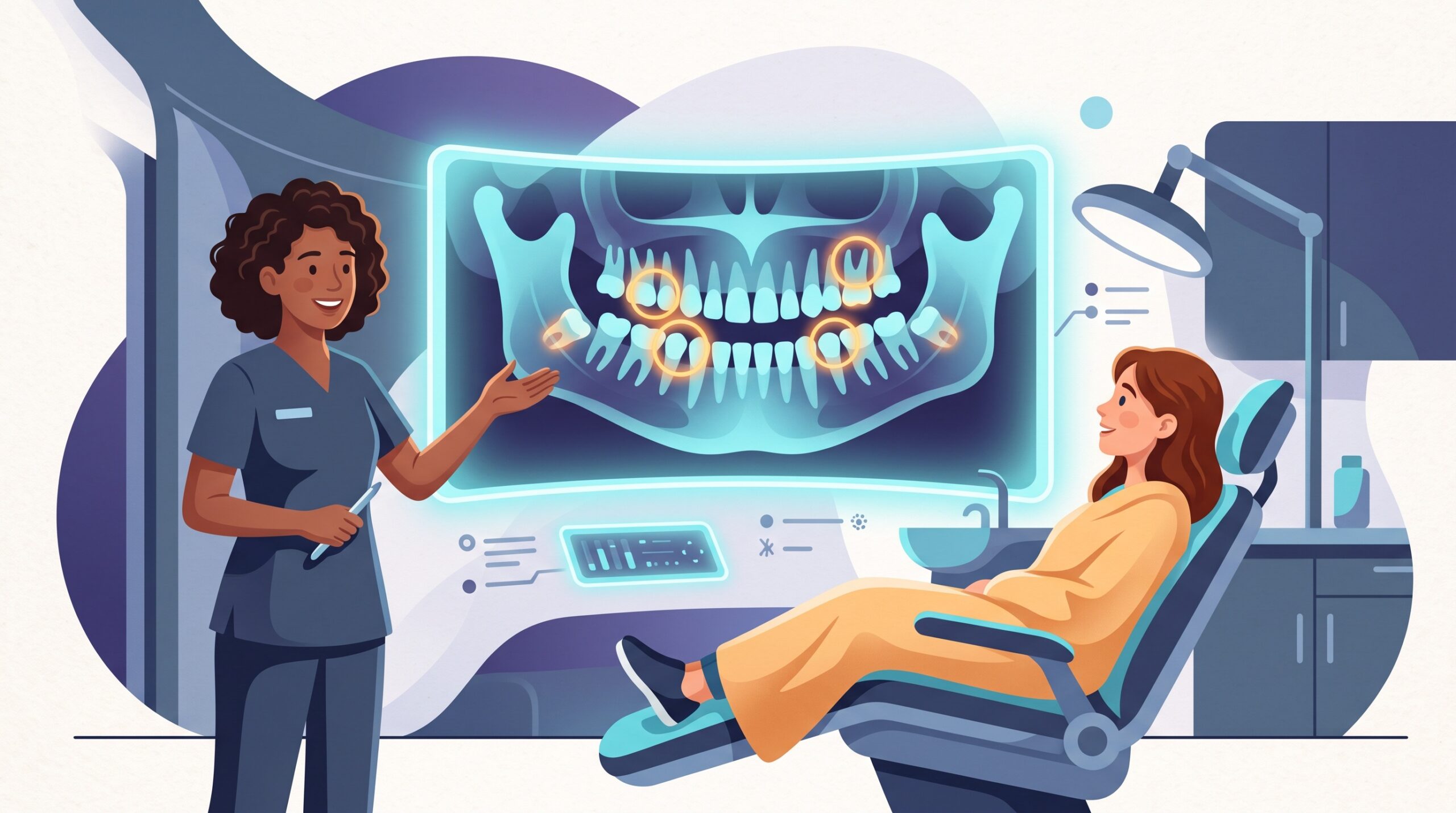
This is the crowded, competitive heart of dental AI, and three names dominate the 2D radiograph space. Overjet is the most feature-heavy: it detects cavities and calculus (that hardened tartar below the gumline), measures bone levels down to the millimeter, and can even automatically clean up a blurry X-ray. Pearl built its reputation on “Second Opinion,” a chairside tool that outlines multiple conditions on bitewing and periapical images the moment they appear. VideaHealth plays in the same arena, known for spotting several categories of findings across a single image.
What they share is more important than what separates them. Each one takes the X-ray your dentist already captured, runs it through a trained model in seconds, and draws attention to spots a human might gloss over. The color-coded overlay you might glimpse on the monitor is not a diagnosis — it is a prompt for a conversation. “The software flagged this; here is what I think.” Done well, that is a genuinely good use of AI: it makes the invisible visible and gives you and the dentist something concrete to look at together.
Going 3D: CBCT and the cloud
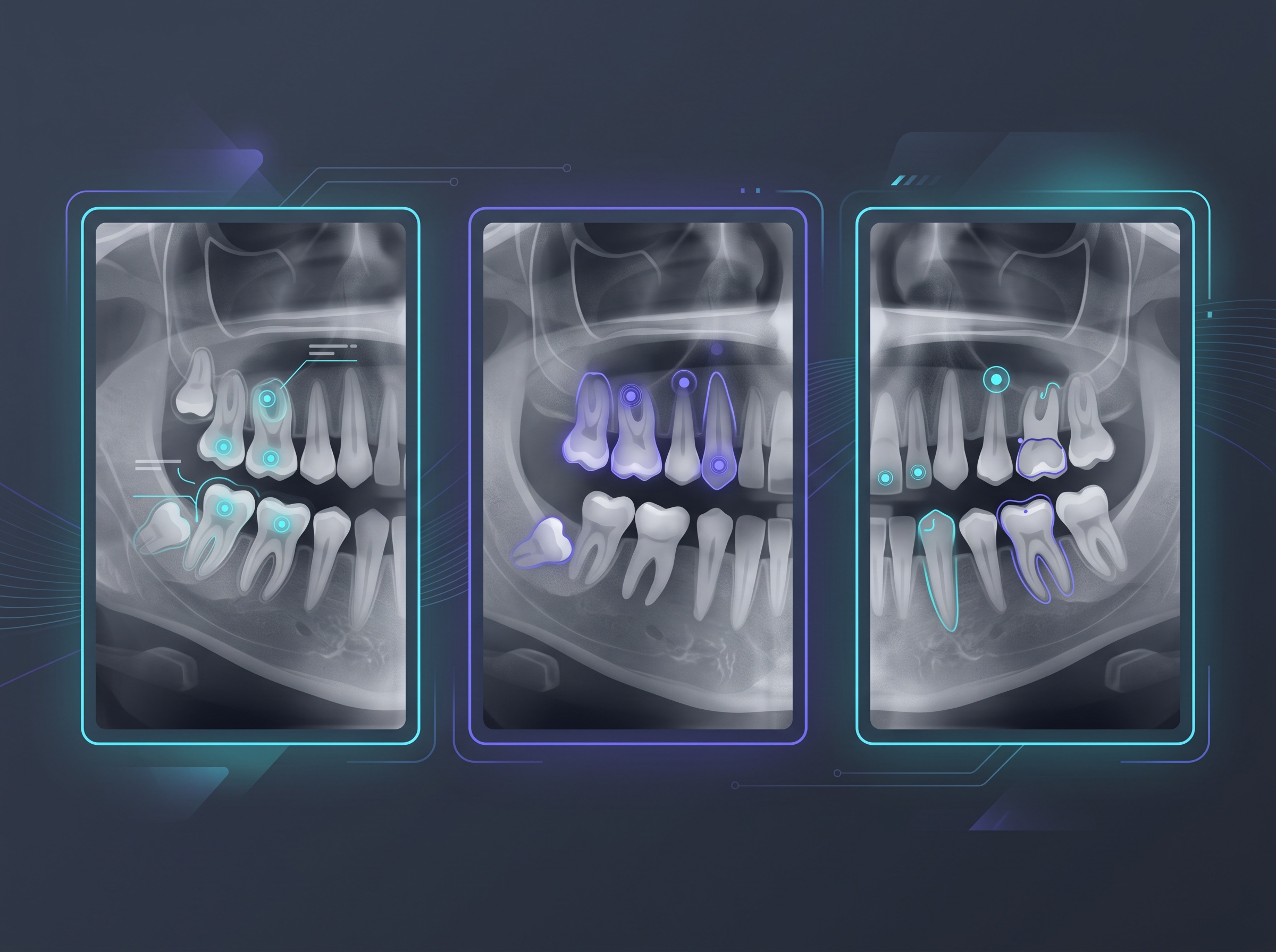
Flat X-rays are only part of modern dentistry. For implants, difficult extractions, and complex cases, dentists use CBCT — a cone-beam CT scan that builds a full 3D volume of your jaw. Reading those is even more demanding, and this is where Diagnocat stands out. It is a cloud-based platform that ingests both 2D X-rays and 3D CBCT scans and returns a structured report: a long list of findings, tooth by tooth, plus automated segmentation that separates individual teeth, nerves, and bone from the 3D data.
The “cloud” part is the quietly significant bit. Instead of every office needing a supercomputer under the front desk, the scan is sent off, analyzed, and returned. That model — capture locally, analyze remotely — is a big part of how dental AI scaled so quickly, and it is a theme we will keep returning to in later articles because it also shapes privacy and integration questions.
Beyond the X-ray: charting, notes, and remote check-ins
Not all dental AI is about imaging. Some of it targets the paperwork that quietly eats a clinician’s day. Denti.AI, for instance, leans into automated charting and AI note-taking — an “AI scribe” that listens during an exam and drafts the clinical notes, so the dentist is looking at you instead of typing. If you have ever watched a doctor stare at a keyboard through your entire appointment, you already understand the appeal.
Then there is remote monitoring. DentalMonitoring uses AI to follow orthodontic cases — clear aligners and braces — from a distance. You take guided photos of your teeth with your phone, the AI assesses whether treatment is tracking as planned, and your orthodontist only pulls you in when something actually needs attention. Fewer pointless check-up drives; the system flags the visits that matter.
“FDA-cleared” — why that phrase matters
You will see “FDA-cleared” attached to these tools a lot, and it is not marketing fluff. It means the software went through a regulatory review before it was allowed to help make clinical calls. As of early 2026, more than a dozen companies hold U.S. clearances for dental AI, and the diagnostic front-runners — Overjet, Pearl, VideaHealth, Denti.AI, and Diagnocat — are among them.
A little nuance worth carrying with you: most of these went through a pathway called 510(k) clearance, which means the tool was shown to be substantially equivalent to something already on the market. A few things travel a different road — DentalMonitoring’s clear-aligner monitoring, for example, came through a De Novo authorization, used when a device is novel enough that there is nothing existing to compare it to. Either way, the point for you is the same: cleared tools have been vetted, and “our AI is FDA-cleared” is a fair question to ask if you are curious whether what is running on your images has cleared that bar. For the regulatory details, the published reviews of FDA-cleared dental imaging AI are refreshingly readable.
What it actually means for you in the chair
So strip away the branding. What changes for a patient? Three honest things. First, a second set of (tireless) eyes on your X-rays, which tends to catch small problems earlier — usually cheaper and less painful to treat than late ones. Second, better conversations: an overlay on the screen makes it far easier for a dentist to show you why they are recommending something instead of just asserting it. Third, a subtle accountability shift — when the same tool reads everyone’s images the same way, there is less room for a rushed or off-day miss.
None of this replaces your dentist’s judgment, and you should be a little wary of anyone who implies it does. The right mental model is a spell-checker, not an autopilot: it catches things, it does not decide things. And it is fair game to ask how it is being used — “Is that the AI flagging it, or you?” is a perfectly reasonable question, and a good dentist will happily answer it.

That is the lay of the land in 2026: AI has stopped being a someday story in dentistry and become a quiet fixture on the operatory monitor. The tools here each deserve a closer look — how they connect to imaging software, what the evidence really says, where the limits are — and that is exactly where this series is headed next. The best way to benefit from any of it is the same as with every good tool: understand what it is actually doing, and let it amplify the human in the room rather than replace them.
Sources & further reading:
- FDA-Approved AI Solutions in Dental Imaging: A Narrative Review (PMC)
- Best AI Tools for Dental Practices in 2026 (AI Dental Tools)
Related Reading
- DentalMonitoring: AI That Follows Ortho Cases From Home
- Overjet, Explained: What Dental AI’s Insurance-and-Imaging Platform Actually Does
- VideaHealth: The AI That Charts Your Teeth Automatically